Published by Beata Boczkowska, Ph.D. on Dec 7, 2023 12:45:17 PM

Apoptosis, a term that describes regulated cell death, is a crucial process in many biological functions such as normal development, maintaining balance, and combating diseases. It is extensively studied due to its significance in understanding the underlying mechanisms of diseases. While apoptosis plays a vital role in the pathogenesis of various conditions, it can become problematic when it occurs excessively in degenerative diseases or insufficiently in others. For instance, in cancer, insufficient activation of apoptosis leads to the persistence and progression of malignant cells. The deficiencies within apoptotic pathways can result in the transformation of affected cells into cancerous ones, tumor metastasis, and resistance to anticancer drugs. Despite its association with cancer development, apoptosis also plays a critical role in cancer treatment as it is a common target for many therapeutic strategies (1).
One therapeutic strategy is by targeting the caspase family of cysteine proteases a well recognized contributor of the apoptotic signaling pathways (1). Caspases are involved in apoptosis and have been categorized based on their mode of operation, specifically as initiator caspases (caspase-8 and caspase-9) or executioner caspases (caspase-3, caspase-6, and caspase-7). For instance, caspase-3 indirectly activates CAD (caspase-activated deoxyribonuclease) while inactivating ICAD (inhibitor of caspase-activated deoxyribonuclease), which contributes to chromatin fragmentation in nucleosome units. Caspases also recognizes several structural proteins as substrates, leading to cleavage that is associated with the distinct morphological changes in apoptotic cells, including chromatin condensation, nucleus fragmentation, and cytoplasmic integrity. Additionally, certain caspases, like caspase-1, play significant roles in signaling pathways related to immune responses against microbial pathogens. In these cases, caspase activation is linked to the maturation of pro-inflammatory cytokines, such as interleukin-1β (IL-1β) and IL-18, rather than apoptosis itself. Therefore it is not surprising that the dysregulation of caspases in turn apoptosis is prominently associated with various human diseases, including cancer, autoimmunity, and neurodegenerative disorders (2).In the field of cancer research, there is a significant focus on the development of new therapeutic strategies focusing on the evasive nature of cancer cells by initiating the apoptosis process (1-2).
Indeed, the detection methods for apoptosis play a pivotal role in apoptosis research and the exploration of therapeutic strategies. The methods should be based on the research objective, necessary to explore and understand the therapeutic drugs and treatment associated with apoptotic diseases. Hence, researchers employ various methods and techniques for detecting apoptosis, including morphology, biochemistry, molecular biology, immunology, and other approaches.
MBL International offers antibodies, recombinant proteins, kits and assays to assist in detection and analysis of different apoptotic molecules. These are some examples of highly cited apoptosis -detection methods using MBLI products.
Enzyme-linked immunosorbent assay (ELISA) is a highly sensitive method that allows for the qualitative and quantitative detection of apoptosis. In this method, the C-terminal ends of the tetrapeptides recognized with by activated caspases are conjugated with p-nitroanilide (pNA) at the C-terminal side and they are used as substrates. After the caspases cleaves the substrates, pNA is released. By the caspase activity can be quantified by measurement of the released emitted fluorescence with a microplate reader, the caspase activities can be quantified (pNA; Abs. 400 nm or 405 nm).

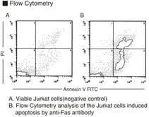
The level of apoptotic cells during the early, middle, and late stages of apoptosis, as well as in necrosis, can be accurately distinguished and quantified using flow cytometry analysis of Annexin V- and PI-stained cells. If the researcher aims to identify the specific location of apoptotic cells, a TUNEL method or related immunofluorescence staining can be employed.
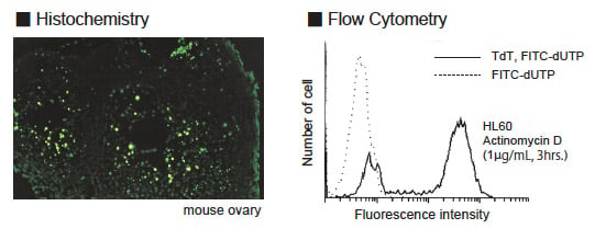
Detection of apoptosis by TUNEL method: Terminal deoxynucleotidyl transferase-mediated dUTP nick-end labeling (TUNEL) method was developed to detect DNA fragmentation in situ. In apoptotic cells, the chromosomal DNA is cleaved by endonucleases at linker DNA sites between nucleosomes. Thereafter, many DNA fragments appear as oligomers of approximately 180 bp in the nuclei. In the TUNEL method, the 3'-OH ends of DNA fragments are nick-end labeled with FITC-dUTP (or dUTP-biotin and avidin-FITC); this process is mediated by terminal deoxynucleotidyl transferase (TdT). TdT catalyzes the addition of deoxynucleotides at the 3'-OH terminus of oligo-deoxyribonucleotide, single-stranded, and double-stranded DNA.
Annexin V Apoptosis Detection Kits
MBLI’s Annexin V apoptosis detection kit is based on the observation that phosphatidylserines (PS) translocate from the inner membrane to the outer membrane during apoptosis. Annexin V binds to PS on the outer membrane. This event can be easily detected using MBLI’s kit using conventional dye-staining along with any streptavidin or avidin-dye reagents.
There are many techniques available for the detection of apoptosis. Researchers may use a combination of techniques to understand apoptotic cells and related targets of endogenous apoptosis pathways both in vivo and in vitro. Regardless of whether you choose to use a single technology or a combination of technologies, it is essential to select the detection method(s) that best align with your research objectives.
References:
-
- Hsu, S. K., Li, C. Y., Lin, I. L., Syue, W. J., Chen, Y. F., Cheng, K. C., ... & Chiu, C. C. (2021). Inflammation-related pyroptosis, a novel programmed cell death pathway, and its crosstalk with immune therapy in cancer treatment. Theranostics, 11(18), 8813.
- Cullen, S. P., and S. J. Martin. "Caspase activation pathways: some recent progress." Cell Death & Differentiation 16.7 (2009): 935-938.


