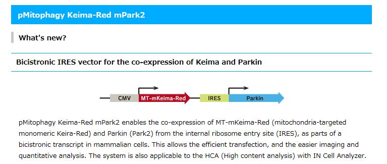
Keima-Red Fluorescent Protein Vector for Mitophagy Monitoring
- Large Stokes shift for clear reporting
- Useful for monitoring mitophagy
- For use with simultaneous multicolor imaging and single laser line FCCS
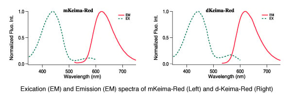
CoralHue® monomeric and dimeric Keima-Red are red fluorescent proteins with the largest commercially available Stokes shift (ex. 440 nm, em. 620 nm) making Keima-Red a superb reporter protein for multicolor fluorescence analysis. Because of this characteristic, they are excited by a very short wavelength but emit a long wavelength. Keima is named after a shogi (Japanese chess) piece Keima that can move in a hopping manner, similar to the knight in the game of chess.
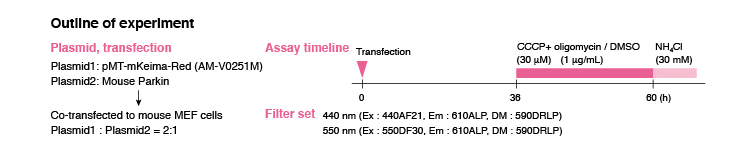
The Keima-Red protein cloning plasmids allow for insertion of cDNA sequences to create protein fusion products between the protein of interest and Keima. Keima-Red cloning plasmids create protein fusion products that are useful for tracking protein localization within cells as well as monitoring gene expression. Keima-Red is also now available as target specific constracts which allow for Keima-Red protein fusion products to be directed to either the mitochondria or the plasma membrane.
Performance and Use
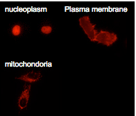
Labeling organelles
dKeima-Red and mKeima-Red are easily expressed in a wide range of organisms. dKeima-Red and mKeima-Red have been demonstrated in the nucleoplasm, plasma membrane, and mitochondria targeting signal models.
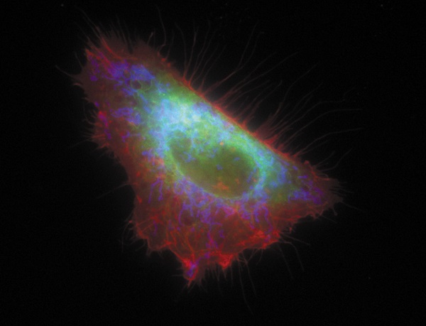
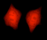
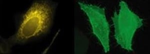
Multicolor labeling of mammalian cells
An image of the HeLa cell labeled with MBLI fluorescent proteins targeting different organelles. mKO1 islocalized on the plasma membrane (red), mAG1 in the endoplasmic reticulum (green) and mKeima-Red in the mitochondria (blue).
The image and information are provided by Dr. Miyawaki Atsushi, Laboratory for Cell Function and Dynamics, BSI, RIKEN
Simultaneous four-color imaging of subcellular structures in a Vero cell using a single laser line (458 nm)
Normalized excitation and emission spectra of SECFP, mMiCy, Venus and mKeima (a). An image of the Vero cell with SECFP localized on the plasma membrane (cyan), mMiCy in the endoplasmic reticulum (green), YFP along the microtubules (red) and mKeima in the mitochondria (purple) (b) . The image was created by merging the following images, which were obtained using spectral imaging: SECFP-pm (c), mMiCy-er (d), Venus-mt (e) and mKeima-mit (f).
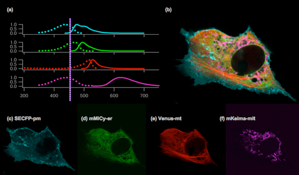
Spectra imaging with a single laser line at 458 nm (Ar ion laser) was performed using the 32 channels of the LSM 510 META system (Carl Zeiss).
Images were kindly provided by Takako Kogure, Atsushi Miyawaki (Laboratory for Cell Function and Dynamics , BSI, RIKEN).
Mitophagy Detection
Keima-Red for mitophagy detection in living cells
- pH dependent excitation profile
- Simple method for creating transfectant cells
- Use with any mammalian cells
- Useful for monitoring mitophagy
Mitophagy is the selective degradation of old or depolarized mitochondria by autophagy and contributes to maintaining a healthy population of mitochondria. Since damaged mitochondria lead to collapse cell homoeostasis, mitophagy is believed to be protective against diseases related to mitochondrial dysfunction such as in neurodegenerative disorders.
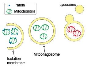
Parkin, an ubiquitin ligase known as the gene responsible for Parkinson’s disease, plays an important role in the autophagic elimination of mitophagy. When mitochondria are depolarized and dysfunctional, PTEN-induced putative kinase protein 1 (PINK1) accumlates on the outer mebrane and recruits Parkin on the damaged mitochondria. The outer membrane on the mitochondria is then ubiquitinated through the ubiquitin ligase activity of Parkin. Finally, the poly-ubiquitinated mitochondria are selectively recognized and executed by the autophagic process.
Emission wavelength character of Keima-Red under various pH condition
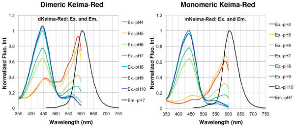
The fluorescent protein Keima has an excitation spectrum that changes according to pH. A short wavelength (440 nm) is predominant for excitation in a neutral environment, whereas a long wavelength (586 nm) is predominant in an acidic environment. The ratio of fluorescent intensity in each excitation condition is an indicator of mitophagy in living cells.
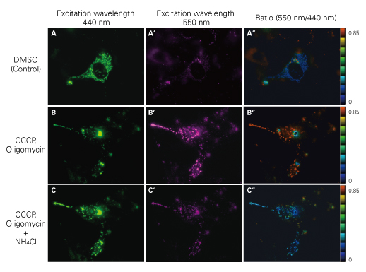
Mitochondrial targeting-mKeima-Red for mitophagy detection
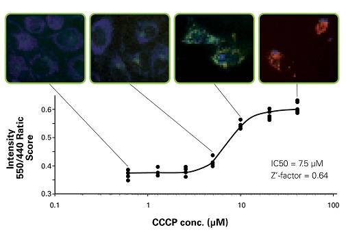
When MT-mKeima-Red, which is the Keima tagged with a mitochondrial-localized signal peptide sequence, and Parkin is expressed in target cells, mitophagy is detected and is visualized through the difference in the fluorescent wavelengths observed before and after drug treatment. Mitophagy is induced by the administration of CCCP and oligomycin, drugs that affect mitochondrial membrane potential and is observed with 550nm/440nm ratio images (A”, B”, C”).
Through mitophagy induction, the mitochondria-localized MT-mKeima-Red is displayed in red in ratio images showing its localization in an acidic environment (B-B”). The neutralizer NH4Cl is then administered which firces the entire cell into a neutral environment and the image turns to blue (C-C”). The result matched findings in which the progression of mitophagy causes mitochondria to be engulfed in lysosomes when in an acidic environment.
Publications: Keima-Red has been referenced in these publications
Citation Highlight
1Bingol B et al. The mitochondrial deubiquitinase USP30 opposes parkin- mediated mitophagy. Nature 510, 370–5 (2014). [PMID: 24896179]
As described in this research paper, living neurons were imaged for the occurrence of mitophagy using Keima-Red. By transfecting vectors into the cells with varying levels of Parkin, PINK1, and USP30, the roles of these proteins in mitophagy were explored. It was found that USP30 opposes Parkin-mediated mitophagy. Since mitophagy has been implicated in Parkinson’s disease, USP30 inhibition might help ameliorate the effects of Parkinson’s by promoting mitochondrial clearance and quality control.
2Katayama H et al. A sensitive and quantitative technique for detecting autophagic events based on lysosomal delivery. Chem Biol. 18, 1042-52 (2011) [PMID: 21867919]
3Choubey V et al. BECN1 is involved in the initiation of mitophagy: It facilitates PARK2 translocation to mitochondria. Autophagy 10, 1105–19 (2014). [PMID: 24879156]
4Safiulina D & Kaasik A. Energetic and Dynamic: How Mitochondria Meet Neuronal Energy Demands. PLoS Biol 11, e1001755 (2013). [PMID: 24391475]
5Togashi K et al. Na+/H+ Exchangers Induce Autophagy in Neurons and Inhibit Polyglutamine-Induced Aggregate Formation. PLoS ONE 8, e81313 (2013). [PMID: 24278418]
6Narendra DP et al. PINK1 rendered temperature sensitive by disease-associated and engineered mutations. Hum Mol Genet. 22, 2572-89. (2013). [PMID: 23459931]
| Code No. | Product | Bacterial Resistance Gene |
|---|---|---|
| AM-V0259M | pMitophagy Keima-Red-Parkin (Kan) | Kanamycin/Neomycin |
| AM-V0259HM | pMitophagy Keima-Red-Parkin (Hyg) | Hygromycin B |
Ratio Imaging
Mitophagy flux is analyzed by the fluorescent intensity ratio of 440 nm and 550 nm. A strong signal at 440nm indicates that mitophagy did not occur while a strong signal at 550 nm indicates mitophagy occured.
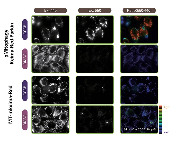
High Content analysis by IN Cell Analyzer
Mitophagy was detected using IN Cell Analyzer 1000. We obtained reasonable IC50 value and a sufficient Z’-factor. pMitophagy Keima-Red -Parkin provides a robust assay for mitophagy monitoring in living cells using high content analyzer.
Learn more about these Related Products
Other Fluorescent Proteins
- Kusabira-Orange: Bright orange fluorescence and high pH stability for gene expression and organelle labeling
- Absorbs light maximally at 548 nm and emits orange light at 561 nm.
- Azami-Green: Bright green fluorescence and high pH stability for protein and organelle labeling
- Absorbs light maximally at 492 nm and emits green light at 505 nm.
- Full Fluorescent Protein Product Listing
Autophagy Antibodies
Our autophagy antibodies are validated in multiple application area (WB, IHC, ICC and IP) and are widely cited, including in research papers with more than 1,000 citations (Saitoh T et al. Loss of the autophagy protein Atg16L1 enhances endotoxin-induced IL-1beta production. Nature 456, 264-8 (2008)).