Published by Dennie Magcase on
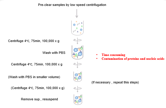
Ultracentrifugation is regarded as the gold standard method for exosome isolation. The ultracentrifugation process is time consuming and may introduce protein and nucleic acid contamination. Pre-clearing samples by one or more low speed centrifugation steps will deplete the cells, platelets, and large apoptotic bodies. Larger extracellular vesicles can be pelleted at forces between 10000-20000 x g. Smaller extracellular vesicles can be pelleted at higher speeds, ranging from 100000-200000 x g. Depending on the protocol, it can take up to 24 hours to isolate exosomes by ultracentrifugation. It is impossible to achieve absolute separation due to potential contamination of proteins and nucleic acids.
Figure 1. General Ultracentrifugation Protocol
ExoCap™ Streptavidin Kit – The faster, easier and customizable
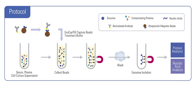
The ExoCap™ Streptavidin kit protocol is very easy and simple to follow. Researchers can isolate and enrich for exosomes of interest by coupling biotinylated molecules to the magnetic Streptavidin beads. There is no need for pre-treatment of samples because there is a direct incubation of sample with the Streptavidin magnetic beads. The magnetic Streptavidin beads coupled with biotinylated antibodies will selectively capture exosomes of interest. During the wash step the captured exosomes on the magnetic bead are retained, while any contaminants are removed, resulting in high purity exosomes that can be used for downstream analyses in as little as 4 hours.
Figure 2. ExoCap™ Streptavidin Kit Protocol
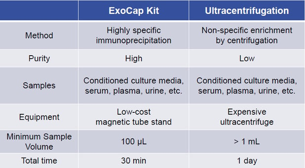
ExoCap™ Streptavidin Kit vs Ultracentrifugation
Table 1. Streptavidin Kit and Ultracentrifugation Comparison