Skip to the main content.

What does mitochondria have to do with mitophagy?

Published by Bindi M. Doshi, PhD on

 What is mitophagy? It is when damaged mitochondria are removed from the cell by autophagy.  The damaged mitochondria end up in lysosomes for their final disposal.  This whole process is to maintain and assure proper cellular function.   The importance of this biological process is that it has been implicated in disease states such as cancer and Parkinson’s disease.

There are two proteins involved with mitophagy, Parkin and PINK1. These proteins have been found mutated in early onset Parkinson’s Disease.  If the pathway for maintaining mitochondria is deficient, the onset of disease occurs.  PINK1 is one of the first to detect if there is a problem with the mitochondrial membrane.  PINK1 will go on to recruit Parkin, which only binds to damaged mitochondria1.  It is thought that Parkin induces mitophagy by ubiquitinating the outer mitochondrial membrane and recruiting other proteins.  At this point, the damaged mitochondria is encompassed by the autophagosome, and subsequently fuses with lysosomes for degradation. 
 
An interesting method for observing mitophagy is by using live cell imaging. MBL has created a vector that combines Keima-Red (fluorescent protein) and Parkin.  Using this one vector, you can now observe mitophagy occurring in real time.  Keima has an excitation spectra that changes with pH.  It changes from 440nm wavelength in a neutral environment to 586nm in an ecidic environment.  Using vector MT-mKeima-Red and drug treatment, one can visualize before and after effects of mitophagy. 
 

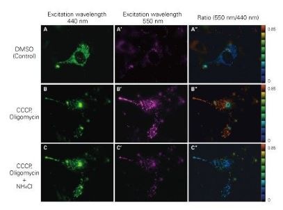
The figure below shows the effects of CCCP and oligomycin, drugs that affect mitochondrial membrane potential. Through mitophagy induction, the mitochondria-localized MT-mKeima-Red is displayed in red in ratio images showing its localization in an acidic environment (B-B”). The neutralizer NH4Cl is then administered, which forces the entire cell into a neutral environment and the image turns to blue (C-C”). The results agree with findings where the progression of mitophagy causes mitochondria to be engulfed in lysosomes when in an acidic environment2.


Find more information for how MBLI can help your mitophagy needs today!


1. Jin, S. M., and R. Youle J. "PINK1- and Parkin-mediated Mitophagy at a Glance." Journal of Cell Science 125.4 (2012): 795-99.  

2. Bingol, Baris, Joy Tea S., Lilian Phu, Mike Reichelt, Corey Bakalarski E., Qinghua Song, Oded Foreman, Donald Kirkpatrick S., and Morgan Sheng. "The Mitochondrial Deubiquitinase USP30 Opposes Parkin-mediated Mitophagy." Nature 510 (2014): 370-375.                                                                                                  


Back to Blog


Comments Or Suggestions