Published by Bindi M. Doshi, PhD on
What are LC3 isoforms all about? What do they do? How are they different? How are they involved in autophagy?
Autophagy is a pretty important cellular process. It allows a cell to survive a state of starvation. What this means is that when a cell is experiencing nutrient poor conditions, autophagy kicks in and basically gets rid of unnecessary debris. You can almost consider it as a necessitated spring cleaning. The cell will only keep “essential” components and remove “non-essential” components. How does this happen? Well, the basic steps of autophagy are: induction (a stimulus will signal to the cell to begin autophagy) and elongation of a double membrane, which is followed by the formation of an autophagosome (material to be degraded is enclosed within the autophagosome membrane). After this point, the autophagosome will fuse with a lysosome- creating an autolysosome (or autophagolysosome). The final step is degradation of the material1. Seems simple enough… until you involve proteins. And LC3 is seriously a gosh darn tricky protein. LC3 is the mammalian homologue of yeast ATG8 (Autophagy related gene 8). Initially, it was characterized as microtubule-associated protein 1 light chain 3 (MAP1LC3)2. LC3 is important because it is considered to be a hallmark for autophagy. So why is this hallmark protein so tricky? Well, LC3 behaves differently in different cell types and for different cell stress3. (Nothing is ever easy, is it?) Despite this, LC3 continues to be widely used in labs for monitoring autophagy.
LC3 has 3 different isoforms: LC3A, LC3B, and LC3C2. There are similarities with the three isoforms as well as differences. All three isoforms show high sequence similarity to rat LC3 according to sequence alignment5. The protein structures of LC3 A, B and C have been determined and all three isoforms show a conserved region required for autophagosome formation6. ATG13 (Autophagy related gene 13) and LC3 are required for autophagosome formation. LC3B is considered to be a popular marker of autophagy. LC3B-I is present in the cytosol. When it is covalently linked with phosphatidylethanolamine (PE), LC3B-I is converted to LC3B-II and is present in the autophagosome membrane during the autophagy cycle4. LC3A and LC3B show different expression patterns in rat tissue implicating there may be functional differences7. The importance and function of each isoform is still under investigation.
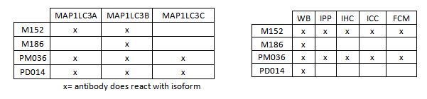
A high quality antibody is especially important when investigating LC3. However, not all antibodies are created equal and finding this high quality antibody can be tricky. Some researchers find a monoclonal anti-LC3 antibody will work better than a polyclonal antibody and vice versa. Much of this success depends on the researcher’s experimental design, cell lines being used, etc. MBL International provides high quality monoclonal and polyclonal LC3 antibodies for use in various applications, including WB, ICC, IHC, IPP, and FCM. A quick reference table is listed below, showing the product code number and the isoform it can detect. An Autophagy Sampler Kit is also available for testing which antibody will provide optimal results for each researcher. For your convenience, as our researchers develop additional LC3 isoform specific antibodies, this blog will be updated!
- Mizushima, N. (2007). Autophagy: Process and function. GENES & DEVELOPMENT, 21, 2861-2873.
- Mann, S., & Hammarback, J. (1994). Molecular characterization of light chain 3. A microtubule binding subunit of MAP1A and MAP1B. Journal of Biological Chemistry, 269(15), 11492-7.
- Klionsky, D. J., Abdalla, F. C., Abeliovich, H., Abraham, R. T., Acevedo-Arozena, A., Adeli, K., … Zuckerbraun, B. (2012). Guidelines for the use and interpretation of assays for monitoring autophagy. Autophagy, 8(4), 445–544. doi:10.4161/auto.19496
- Barth, S., Glick, D., & Macleod, K. (2010). Autophagy: Assays and artifacts. The Journal of Pathology, 221(2), 117-124.
- He, H. (2003). Post-translational Modifications of Three Members of the Human MAP1LC3 Family and Detection of a Novel Type of Modification for MAP1LC3B. Journal of Biological Chemistry, 278, 29278-29287.
- Suzuki, H., Tabata, K., Morita, E., Kawasaki, M., Kato, R., Dobson, R., . . . Wakatsuki, S. (2014). Structural Basis of the Autophagy-Related LC3/Atg13 LIR Complex: Recognition and Interaction Mechanism. Structure, 22(1), 47-58.
- Wu, J., Dang, Y., Su, W., Liu, C., Ma, H., Shan, Y., . . . Yu, L. (2005). Molecular cloning and characterization of rat LC3A and LC3B—Two novel markers of autophagosome. Biochemical and Biophysical Research Communications, 339(1), 437-442.