
Published by M. Mamunur Rahman, PhD on

Cell-based assays are the first step for drug screening applications and have been performed in monolayer culture condition for decades. These traditional cell cultures have been known to enhance integrin signals, which mask several biologic activities in tumor cells. In contrast, it is already known that 3D cell culture can replicate the intra-tumor microenvironment.
Epithelial Mesenchymal Transition (EMT) is a biologic process for carcinoma progression as it is related to cancer invasion or metastasis that remains the primary cause of cancer mortality worldwide. In EMT process, tumor cells dissociate and become motile, leading to localized invasion and metastatic spread. In addition to the morphological changes, key factors/markers in the steps of EMT have been established including cell adhesion molecules and, transcription factors and kinases[i]. During cancer progression, cancer cells assume a spindled shape, lose desmosomes and adherence junctions, and express mesenchymal genes, such as vimentin and zeb-19,[ii]. These EMT markers are correlated with drug resistance[iii] and tumor progressions[iv]. Targeting EMT therefore represents an important therapeutic strategy for cancer treatment. Recently, Kumar et al. reported that mesenchymal transition of non-small cell lung cancer (NSCLC) cell lines were much more efficiently induced on 3D cell culture than that on 2D cell culture[v]. This result persuades us to develop a novel 3D screening system.
In our recent publication Arai et al.[vi], we have introduced an innovative three-dimensional (3D) cell based high throughput screening assay that can lead to the identification of EMT inhibitors. We have cultured cells on NanoCulture Plate (NCP), a micro-patterned gel free scaffold type 3D cell culture plate, to mimic in vivo tumor micro environment. This ECM mimicking structure on the NCPs restricts cells to sprawl on the base and enable tumor cells to migrate more than monolayer cell culture. Cells can migrate more actively from a scaffold to the other scaffold on the grid in the NCPs than cells on 2D cell culture plate, on which cells migrate less. On the other hand, easy handling, microscopic compatibility, in plate application performance, and SBS format compatibility make NCP better platform than other scaffold based 3D system. Therefore the NanoCulture Plates are useful for scaffold-based type 3D tissue culture system based on micro-patterned surface without gels.
Lung cancer cell lines have been treated with TGFβ to stimulate the spheroid to induce EMT and observed the extent of EMT by the morphology change under a HTS measurement method using hypoxia probe, in order to quantify more accurate data. This “add only” assay is simplest and suitable for HTS. Further researchers may enable to cross validate their data performing WB/RNA of common EMT biomarkers panel (i.e. Vimentin, Zeb-1, N-cadherin, E-cadherin) at the end as this is a live cell imaging probe. A pilot screen has been conducted to validate this model for HTS using LOPAC and identified several compounds as viable EMT inhibitors. Overall, we hope that this 3D EMT HTS method will provide an immense improvement of therapeutic medicine.
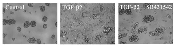
Fig: EMT on NanoCulture Plate. A549 cells were cultured with or without TGF-β2 only or TGF-β2 + SB431542 for 4 days. Representative images were shown. Scale bar, 200 μm
-----------------------------------------------------
[i] Lamouille S, Xu J, & Derynck R. Molecular mechanisms of epithelial–mesenchymal transition. (2014) Nature reviews Molecular cell biology. 15(3): 178-196.
[ii] Wellner U, Schubert J, Burk UC, Schmalhofer O, Zhu F, Sonntag A, Waldvogel B, Vannier C, Darling D, zur Hausen A, Brunton VG, Morton J, Sansom O, Schüler J, Stemmler MP, Herzberger C, Hopt U, Keck T, Brabletz S, Brabletz T. The EMT-activator ZEB1 promotes tumorigenicity by repressing stemness-inhibiting microRNAs. (2009) Nat Cell Biol. 11: 1487-95.
[iii] Pan JJ, Yang MH. (2011) The role of epithelial-mesenchymal transition in pancreatic cancer. J Gastrointest Oncol. 2: 151-6.
[iv] Liu J, Chen L, Deng H, Xu B, Li M, Zheng X, ... & Jiang J. Epithelial-to-mesenchymal transition in human esophageal cancer associates with tumor progression and patient’s survival. (2014) International journal of clinical and experimental pathology. 7(10): 6943.
[v] Kumar M, Allison DF, Baranova NN, Wamsley JJ, Katz AJ, Bekiranov S, Jones DR, Mayo MW. NF-κB regulates mesenchymal transition for the induction of non-small cell lung cancer initiating cells. (2013) PLoS One. 8: e68597.
[vi] Arai K, Eguchi T, Rahman MM, Sakamoto R, Masuda N, Natatsura T, Calderwood SK, Kozaki KI, Itoh M. A Novel High-Throughput 3D Screening System for EMT Inhibitors: A Pilot Screening Discovered the EMT Inhibitory Activity of CDK2 Inhibitor SU9516. (2016) PLoS One. 11(9): e0162394.